

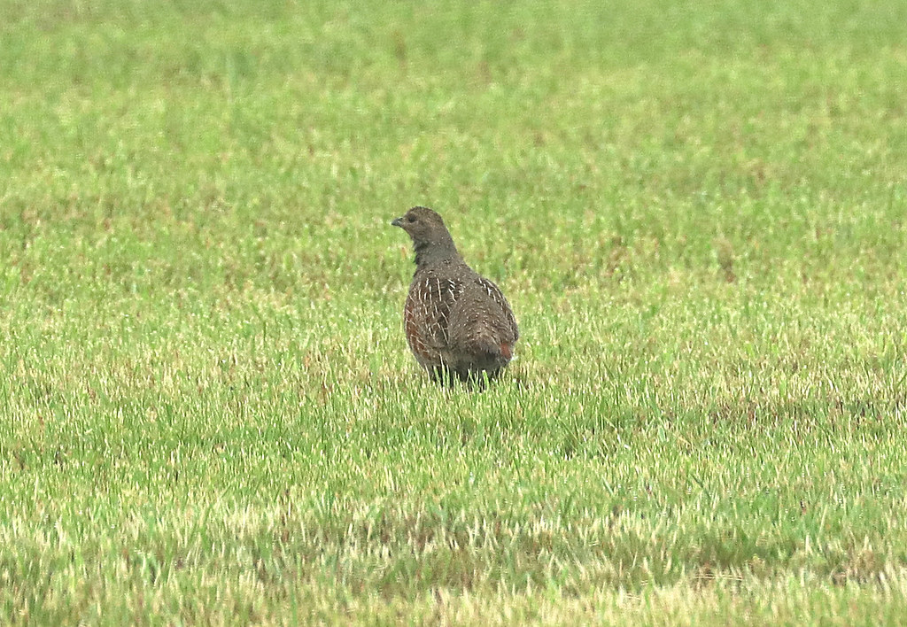
Coccidia lifecycle
Coccidiosis is considered the single most common disease in gamebird production, this intestinal parasite is transmitted in faeces by single celled organisms called oocysts. The production and spread of this parasite is very fast, one oocyst can reproduce resulting in millions of oocysts. If left undetected coccidia can cause waves of very ill birds and numerous deaths. The effects it may have on partridges are explored in this article.
When oocysts are within the faeces or environment moisture and warmth causes them to sporulate making them “infective”. The birds then ingest these oocysts and the cycle within the intestinal tract starts, multiplying and causing illness. Depending on the coccidia species, after approximately 3 days the oocysts are then emitted through faeces and the cycle starts again. Numerous different coccidia species can be found in each bird, these usually affect different areas of the intestinal tract. If you are suffering from coccidiosis in your flock, contact a veterinarian for assistance. For more on sources of coccidiosis, read here.
Coccidia species affecting partridges and their signs
The most common coccidia species found in partridges reared in the UK are: Eimeria kofoidi, Eimeria caucasica and Eimeria legionesis.
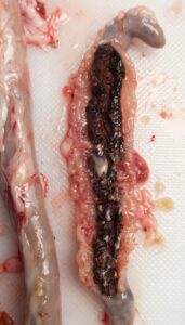
Figure 1: Pictured is a full caecal core formed from the blood produced by lesions in the digestive tract.
Eimeria kofoidi: This species can cause partridges to become weak and emaciated. The prepatent period is 5
days. The parasite starts in the duodenum and jejunum, however heavy infections cause the parasite to spread to the ileum. Commonly the duodenum and jejunum appear white, dilated and swollen. White spots can be seen from the outside of the intestine, the duodenum contains think mucus and white streaks, thick green or yellow mucus is found in the upper intestine.
The oocysts are identified by being spherical to broadly ovoid, brown or yellowish in colour with slight blueish colour on the inside. They have no micropyle and no oocyst residuum but have numerous small polar granules.
Clinical signs include mucus in faecal droppings, huddling, drooping wings, ruffled feathers, halved closed eyes, depression, loss of appetite and reduced weight gain.
Eimeria caucasica: This species can cause partridges to become weak and emaciated. The prepatent period is 5 days. The parasite invades the duodenum and jejunum similar to that of E. kofoidi, the main difference between the two species being the lesions spotted from the outside of the intestine which are pinkish/reddish spots for E. caucasica. Mucus with the colour of orange/yellow can be found in the duodenum, eventually the intestine becomes very pale and fragile.
The oocysts are identified by being ellipsoid/ovoid with no micropyle. They have a yellowish/blue/green bilayered wall and they have one large granule but no oocyst residuum.
Clinical signs include huddling, drooping wings, ruffled feathers, halved closed eyes, depression, and loss of appetite and reduced weight gain.
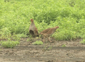
Figure 2: Two grey Partridges (Perdix perdix), Native to the UK.
Eimeria legionesis: This species can cause partridges to become weak and emaciated. The prepatent period is 5 days. This parasite mainly invades the caecae of the bird’s intestinal tract. Petechial hemorrhaging is common inside the folds of the caecae with no normal caecal contents present, instead there are cheesy white cores sometimes in fragments but with heavy infections it is seen as one whole core. Most frequently only one caecae is affected by cores, the other usually has yellowish frothy contents.
The oocysts are identified by having a smooth curve with symmetrical flattened walls, they were smooth, bilayered, yellow/brownish on the outside and blue/greenish on the inside. They possess a micropyle with one or two granules, no oocyst residuum present.
Clinical signs include white caecae core fragments found in droppings, reddish droppings, huddling, drooping wings, ruffled feathers, halved closed eyes, depression, loss of appetite and reduced weight gain.
Identifying coccidia species is a service we provide here at Ridgeway Research Ltd called speciation. We do this by extracting the oocysts out of the infected faeces, incubating them to provide them the warmth to sporulate, then identify the characteristics under a microscope. The lesions that are caused by coccidia can be used to help confirm an identity of a species.
It has been found that the red leg partridge are more susceptible and sensitive to a coccidia infection so sudden deaths are not uncommon for this species.
Find this interesting? Read about coccidiosis in pheasants here.
Please email lab@ridgewayresearch.co.uk for any questions you may have.