

Coccidia lifecycle
Coccidiosis is considered the single most common disease in gamebird production, this intestinal parasite is transmitted in faeces by single celled organisms called oocysts. The production and spread of this parasite is very fast, one oocyst can reproduce resulting in millions of oocysts. If left undetected coccidia can cause waves of very ill birds and numerous deaths. The effects it may have on pheasants are explored in this article.
When oocysts are within the faeces or environment moisture and warmth causes them to sporulate making them “infective”. The birds then ingest these oocysts and the cycle within the intestinal tract starts, multiplying and causing illness. Depending on the coccidia species, after approximately 3 days the oocysts are then emitted through faeces and the cycle starts again. Numerous different coccidia species can be found in each bird, these usually affect different areas of the intestinal tract. If you are suffering from coccidiosis in your flock, contact a veterinarian for assistance. For more on sources of coccidiosis, read here.
Pheasant coccidia species and their lifestyles
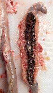
Figure 1: Pictured is a full caecal core formed from the blood produced by lesions in the digestive tract.
The most common coccidia species found in pheasants reared in the UK are: Eimeria colchici, Eimeria duodenalis and Eimeria phasiani.
Eimeria colchici: This species is the most pathogenic found in pheasants, causing weight loss and mortality in infected birds. The oocysts typically sporulate in 2 days and the prepatent period (time between ingestion and excretion) is approximately 6 days. The parasite starts in the small intestine of the bird then eventually moves to the caecae, here it causes white cores and bloody contents. Scrapings of the bird’s caecae and lower intestine will show high numbers of oocysts present.
The oocysts are identified by being elongated, ellipsoidal with one side less rounded than the other and colourless. They include a discreet micropyle, a polar granule but no oocyst residuum.
Clinical signs of this species include diarrhea and white soiling around the vent.
 Eimeria duodenalis: This species is less pathogenic than E. colchici, however can still cause mucoid enteritis in the upper small intestine of birds. The oocysts typically sporulate in 1 to 2 days and the prepatent period is approximately 5 days. The parasite starts in the duodenum of the birds intestinal tract then spreads to the upper small intestine, in highly infected birds the parasite may spread to the whole intestine. Mucoid enteritis in the duodenum is a sign of E. duodenalis, however scrapings of this area and upper small intestine will show a number of small oocysts. The intestine may be blocked and contain a pinkish mucoid exudate, the caecae may be swelled with a foamy yellowish fluid.
The oocysts are identified by being sub spherical to broadly ellipsoidal and smooth. They can be colourless but also have a slight pale yellowish to brown colouring. They have no micropyle and no oocysts residuum.
Clinical signs of this species are to harder identify because light infections may show no signs but heavy infections are identified by the appearance of mucoid enteritis.
Eimeria phasiani: Mortality in birds infected with this species can reach 50% in 2 to 3 week old pheasants. The oocysts typically sporulate in 2 days and the prepatent period is approximately 5 days. The parasite starts in the small intestine but can spread as far as the upper small intestine into the duodenum and lower small intestine into the caecae. Similar to E. duodenalis, mucoid enteritis is a typical sign, however scrapings of the small intestine and proximal part of the caecae will show oocysts. Heavy infections show petechial hemorrhaging, blood-streaked mucus and ballooning throughout the infected intestine.
The oocysts are identified by being ellipsoidal, smooth and yellowish in colour. They have no micropyle and no oocyst residuum but do contain one to three polar granules.
Clinical signs of this species include anorexia, depression and a reduced weight gain in birds. During heavy infections the faeces may be liquid with mucus present and small amounts of blood.
Find this interesting? Read about coccidiosis in partridges here.
Please email lab@ridgewayresearch.co.uk for any questions you may have.